Welcome to 广东脊祥万岁健康管理有限公司!
Service hotline:
13533607738
18602592233
Welcome to 广东脊祥万岁健康管理有限公司!
Service hotline:
13533607738
18602592233

最新资讯
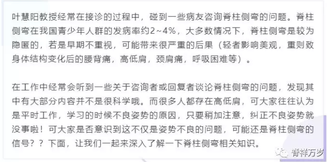
The causes of scoliosis are very complex and related to other factors. Classic summary: the relationship between laying hens and laying eggs
Therefore, early detection and early treatment should be carried out.
Early screening
In the process of formation and development of scoliosis, it is easy to be neglected because of little pain or discomfort, and adolescents have strong self-care ability. Parents and children themselves are not easy to detect, and even form structural scoliosis of the spine. For this reason, the importance of regular screening can be imagined!!! It is generally recommended that regular screening be started every three months at the age of 8.
Parents should pay attention to the development of their children's spine to see if there are the following problems:
(1)Two shoulder uneven
(2)Unequal height of scapula on both sides
(3)Lumbar asymmetry
(4)Hip lifting
(5)Back asymmetry on both sides during forward bending
If you have any of the above problems, you should see a doctor immediately!
Diagnostic evaluation
(1)General history
(2)History of operation
(3)Time of deformity
(4)History of back pain
(5)Cardiopulmonary function
(6)family history
Scoliosis has obvious features in appearance and posture. We can evaluate it by observing its appearance.
(1)The range of motion of the joints with flexion, extension, lateral flexion and rotation of the spine is partially limited;
(2)Shoulders unequal and asymmetrical;
(3)Hexa unequal height, lower limbs unequal length;
(4)Trunk tilt, back asymmetry;
(5)Asymmetric breast development, razor back deformity, etc.。
The examinee exposed the spine as far as possible, stood with feet together, knees straight, arms hanging naturally, trunk bending forward to the back close to the level; the examiner observed the abnormal changes of the spine and thorax along the horizontal plane behind him.
General forward bending test can find the change of back height on both sides. If one side of the ridge appears, it shows that there must be rotation deformity in the rib and vertebral body. Further measurement of rotation angle is needed.
The height of the protuberance can be measured with a level gauge, and the rotation of the trunk can be measured with a square goniometer and a scoliometer. Generally speaking, the greater the height difference between bilateral rib bulges, the greater the angle of lateral bending. Of course, the above measurement of rotation over 4 degrees is considered to be a problem.
X-ray is especially important, it is the true and reliable evidence of scoliosis. The gold standard of diagnosis is often that scoliosis is unreliable without X-ray. We can use X-ray film to determine the type and severity of spinal deformity, understand the etiology, help to select treatment methods and judge the curative effect.
1)Cobb angle measurement
This Cobb method is the most commonly used method to measure scoliosis angle; it must be measured by professionals, because if the selection of vertebral body is incorrect, it will cause the deviation of measurement angle, and mistakenly fasten the cap to others!
(1)Key Points for Reading Films:
A.End vertebrae: the body at the top and tail end of the bend;
B.Acrospinal vertebra: The vertebral body that rotates most severely in bending and deviates most from the perpendicular line;
C.Main side bending (i.e. primary side bending): The earliest bending is the largest structural bending, with poor flexibility and correctability.
D.次侧弯(即代偿Sexual or secondary scoliosis: The smallest bend, which is more elastic than the main scoliosis, but structural or non-structural, is located above or below the main scoliosis; the function is to maintain the normal force line of the body, and the vertebral body usually does not rotate.
(2)Cobb horn:
The angle between the perpendicular line of the lengthening line at the upper edge of the head and the perpendicular line at the lower edge of the caudal vertebra. It is not only suitable for the diagnosis before treatment, but also for the evaluation of curative effect after treatment. (Cobb angle less than 10 degrees is negative, 10-20 degrees is positive, more than 20 degrees is obviously positive)
2)Vertebral rotation measurement (Nash-Moe method)
There are many measurement methods, but Nash-Moe method is the most commonly used method in clinical measurement of scoliosis vertebral rotation.。
(1)Main points:
The standard of measuring the change of the position of the convex and concave pedicle in the ortho-position X-ray film was used to evaluate the degree of vertebral rotation, which was divided into five grades. On the orthopaedic radiographs, the vertebral body was divided into 6 segments, with 1-6 segments from convex to concave sides.
(2)Rotation Level Five Assessment:
Grade 0: (no rotation), bilateral pedicles are in normal, symmetrical position, located in the lateral segment.
Grade I: Convex pedicle slightly moved inward, but still in the lateral segment; concave pedicle shifted outward, and the peripheral image gradually disappeared.
Grade II: Convex pedicle image moved to the second segment; concave pedicle disappeared basically.
Grade III: Convex pedicle images were moved to the midline of the vertebral body or in the third segment; concave pedicle disappeared completely.
Grade IV: The convex pedicle crosses the midline to the fourth segment and is located on the concave side of the vertebral body. The concave pedicle disappears completely.
<br style="margin: 0px; padding: 0px; max-width: 100%; box-sizing: borde

CopyRight © 2017 广东脊祥万岁健康管理有限公司 All Rights Reserved 苏ICP备15053449号-1