Welcome to 广东脊祥万岁健康管理有限公司!
Service hotline:
13533607738
18602592233
Welcome to 广东脊祥万岁健康管理有限公司!
Service hotline:
13533607738
18602592233


最新资讯
There are two main sources of thrombosis in cerebral infarction caused by blockage of cerebrovascular:
First source,
Cerebrovascular thrombosis
All the arteries and blood vessels in the human body can have long plaques, and the blood vessels in the brain are no exception. After the plaque rupture, thrombus quickly formed and blocked the blood vessels.
Second source,
Thrombosis in other parts of the body: Like floating wood in a river, thrombus can flow around the body with blood.
The blood vessel is dense and thin, and other blood vessels can flow past the thrombus, which is blocked in the fine blood vessel.
There are two main sources of such thrombosis:
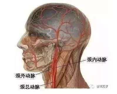
Blood in the brain flows from the carotid and vertebral arteries.
Arteries accounted for 80% and vertebral arteries 20%.
The carotid artery is very close to the brain and can be touched on both sides of the neck. It is divided into common carotid artery, internal carotid artery and external carotid artery.
The internal artery is divided into anterior cerebral artery and middle cerebral artery.……。
The carotid artery is one of the three most vulnerable places in the body to long plaques.
Carotid artery has bifurcation, where the bifurcation is strongly impacted by blood flow, which is most prone to long plaque of vascular endothelial rupture.
The larger the patch, the more fragile it is.
It's like mud that's stuck on the wall but not solid.,
Thrombosis occurs when the blood flow is impacted and falls off.
Once the thrombus is formed, it will immediately flow into the brain and cause cerebral infarction.
Have you heard the word "atrial fibrillation"? "Atrial fibrillation" is short for "atrial fibrillation", is a very common serious arrhythmia. Normally, the heart pumps blood into the aorta smoothly through regular contractions to circulate blood.
When atrial fibrillation occurs, the heart can't contract normally and effectively. It can only tremble quickly and uncoordinatedly, which leads to the decline of blood pumping function. The blood pump can't go out and can only be deposited in the heart. This condition is very easy to form thrombus on the inner wall of the atrium, which is called "mural thrombus".
These thrombi circulate throughout the body.,
Flow to the brain will block up the cerebrovascular, leading to cerebral infarction;
Flow to the lower extremity will block the lower extremity arteries, leading to amputation.
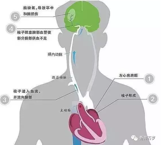
The possibility of cerebral infarction caused by atrial fibrillation Increase 5 times
20% cerebral infarction It is caused by atrial fibrillation.
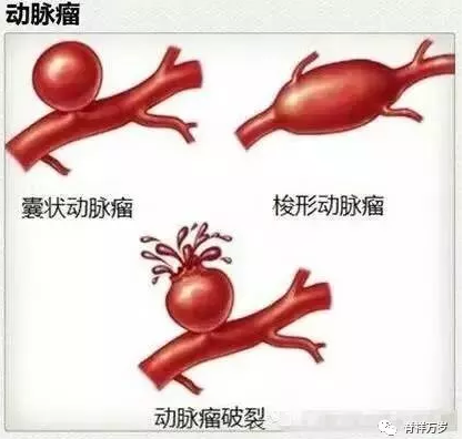
The blood vessel is very thin and its wall is very thin. After atherosclerosis, the cerebrovascular is like the aging "bicycle inner tire". When the pressure is slightly higher, it bulges up a bag. This bag is a microaneurysm. There are a lot of such microaneurysms in the atherosclerotic cerebrovascular. At the same blood pressure, these places are equivalent to being "blown up", the blood vessel wall is thinner, so it is easier to rupture and cause cerebral hemorrhage.
Moreover, the cerebral artery and its branches are mostly at right angles, and the bifurcation should bear greater impact of blood flow, which is also an important reason for the easy rupture of cerebrovascular.
The severity of cerebral hemorrhage depends on the location and amount of hemorrhage.
Figure: A represents subcortical hemorrhage; B represents basal ganglia hemorrhage; C represents thalamic hemorrhage; D represents brainstem hemorrhage; E represents cerebellar hemorrhage.
①Basal ganglia hemorrhage:
Putamen: the most common bleeding site, when the amount of bleeding is small, it causes the opposite side of the body hemiplegia, sensory impairment, binocular gaze to the side of the lesion, etc. when the amount of bleeding is large, the patient quickly coma.
Thalamus: Contralateral limb paralysis (lower limbs are more severe than upper limbs), sensory disorders, language disorders, mental disorders (such as apathy, depression), gaze at the tip of the nose, pupil shrinkage, high fever, etc.
②Brainstem hemorrhage:
Brain bridge: When the amount of bleeding is small, it is characterized by sudden headache, vomiting, vertigo, diplopia and quadriplegia; when the amount of bleeding is large, the patient will soon develop consciousness disorder, needle-like pupil, quadriplegia, dyspnea and high fever, and often die within 48 hours.
Midbrain: Sudden diplopia, blepharoptosis, pupil enlargement, ipsilateral limb ataxia in mild cases, and rapid death in severe cases.
Medulla oblongata: sudden collapse, disturbance of consciousness, decrease of blood pressure, irregular breathing, arrhythmia of heart rate and death.
③Cerebellar hemorrhage:
When a small amount of bleeding occurs, it manifests as nystagmus, ataxia of lesion side, instability of standing and gait, rigidity of neck, etc. When a large amount of bleeding occurs, the patient will soon coma, the pupil shrinks like a needle tip, irregular breathing and death.
④Subarachnoid hemorrhage:
It is caused by the rupture of blood vessels at the base of the brain or on the surface of the brain and the flow of blood into the subarachnoid space. Patients with sudden severe headache (explosive pain, intolerable, persistent can not alleviate or aggravate), often accompanied by nausea, vomiting, can be conscious disorders, delirium, hallucination and other mental symptoms, a small number of epileptic seizures.
In short, the brain is the most sophisticated organ of the human body, there are innumerable blood vessels in it, which can not be a problem!
How strong is the blood vessel and how long is its life span?

CopyRight © 2017 广东脊祥万岁健康管理有限公司 All Rights Reserved 苏ICP备15053449号-1