Welcome to 广东脊祥万岁健康管理有限公司!
Service hotline:
13533607738
18602592233
Welcome to 广东脊祥万岁健康管理有限公司!
Service hotline:
13533607738
18602592233


最新资讯
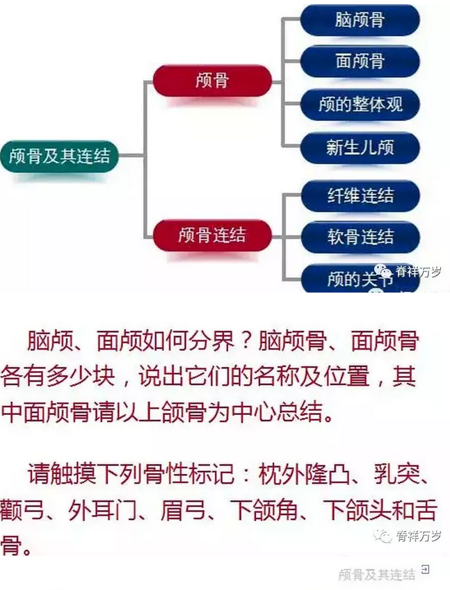
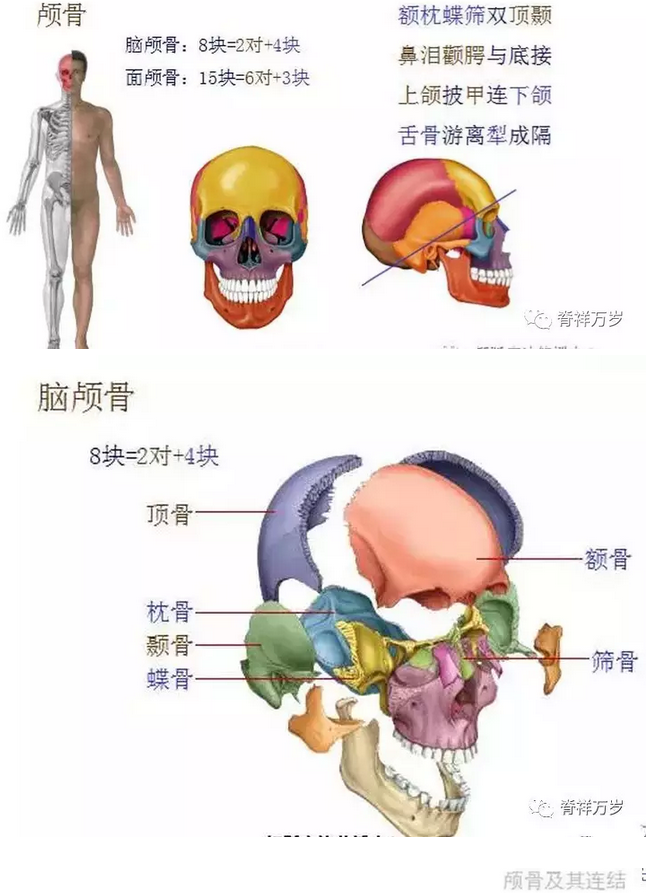
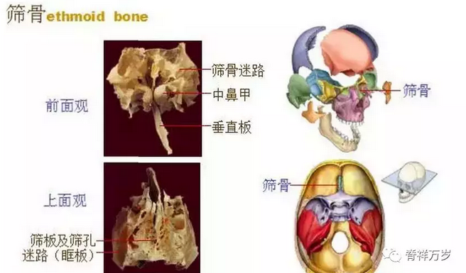
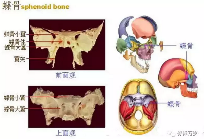
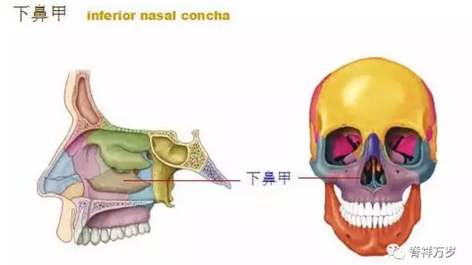
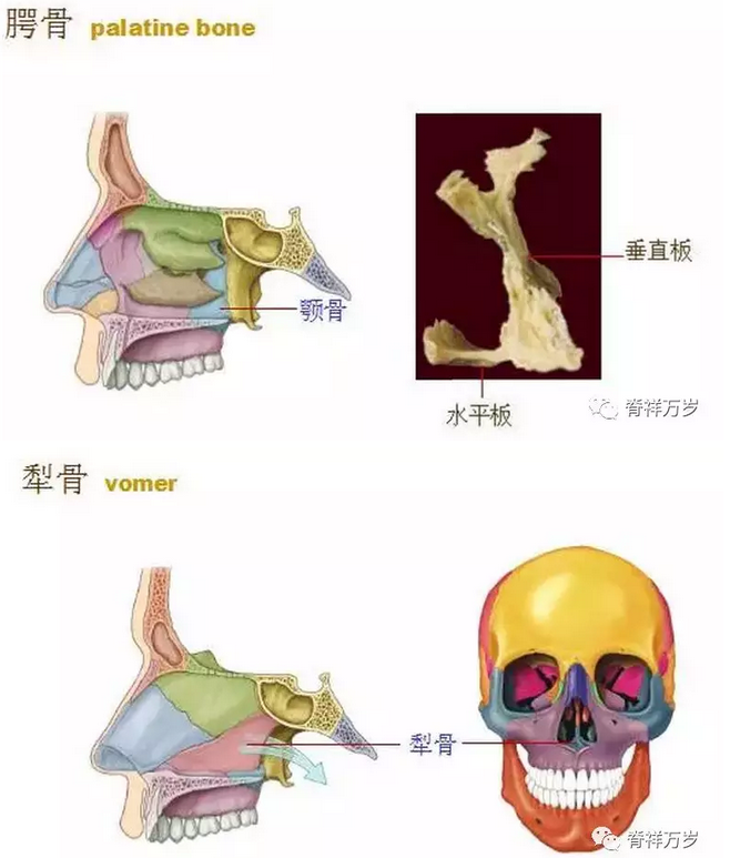
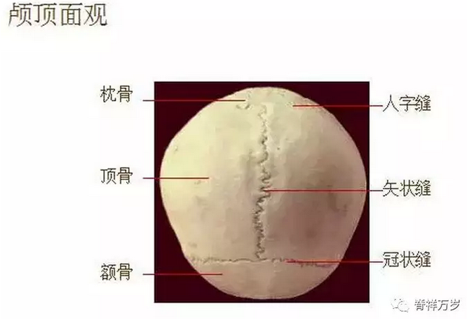
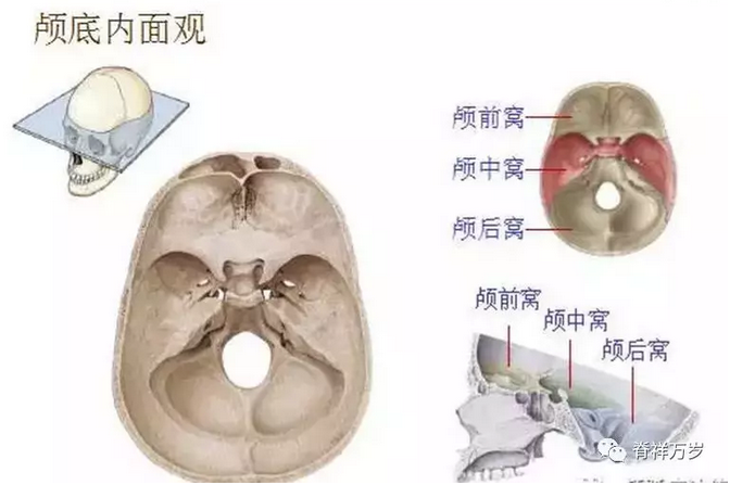
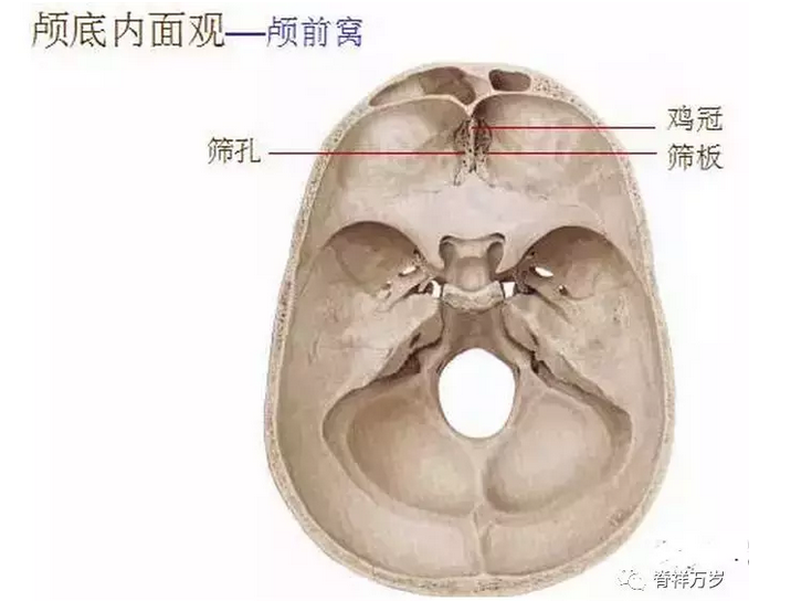
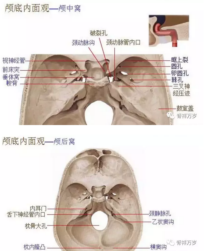
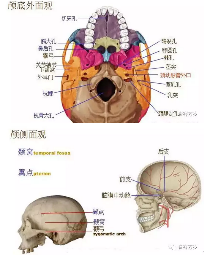
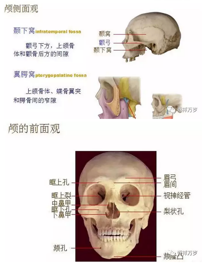
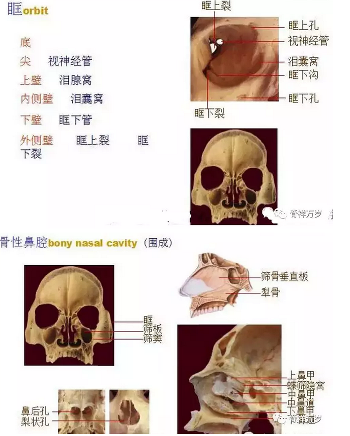
1. On the top of the skull, it is oval in shape, wide in front and narrow in back. The upper part of the skull is called the skull cap. There are three sutures: the coronal suture between frontal bone and parietal bone, the sagittal suture between parietal bone and the herringbone suture between parietal bone and occipital bone. 2. Cranial lateral view consists of frontal bone, sphenoid bone, parietal bone, temporal bone and occipital bone. In the middle of the side, there is the external ear door. The protuberance behind the external ear door is the mastoid process. In the temporal fossa, there are pterions at the junction of frontal bone, parietal bone, temporal bone and sphenoid bone. 3. Precranial view: orbital and osseous nasal cavity. (1) The orbit is divided into bottom, apex and four walls. The orbital apex has optic nerve foramen, and the inferior orbital wall has infraorbital groove, canal and foramen. (2) The lateral wall of the skeletal nasal cavity has three bone slices protruding downward, which are called upper turbinate, middle turbinate and inferior turbinate respectively from top to bottom. The space under each turbinate is called upper nasal meatus, middle nasal meatus and lower nasal meatus respectively. 4. Outside the skull base, the front part of the skull base is called the palate, which is surrounded by the horizontal plate of the maxilla and palate. The middle part is the pterygoid process of the sphenoid bone. In the middle part, there is a large foramen, which is called the foramen occipitalis. There are rupture foramen, jugular foramen and external orifice of the carotid artery canal in the front and 5. The intracranial view of the skull base is divided into three fossa from front to back. (1) anterior cranial fossa consists of frontal orbital part, ethmoid plate and sphenoid wing. (2) The middle cranial fossa consists of sphenoid body, great wing and petrous part of temporal bone. (3) Posterior fossa occipital foramen magnum, occipital protuberance, transverse sinus sulcus, sigmoid sinus sulcus and hypoglossal nerve canal.


The information comes from the Internet and the copyright belongs to the original author. If there is any infringement, please contact us in time and we will delete it. (If there are similarities, the right of interpretation belongs to Long Live Jixiang)

CopyRight © 2017 广东脊祥万岁健康管理有限公司 All Rights Reserved 苏ICP备15053449号-1